“A NEW APPROACH TO RADIAL NERVE PALSY IN CATS”. CLINICAL CASE SERIES REPORT

- Introduction
The radial nerve palsy is a pathology that is rarely seen in dogs, in comparison to cats, where it is more commonly seen, especially in young stray cats. The most commonly observed clinical picture in such patients includes paralysis of the antebrachial portion of the limb, the carpus, the manus and fingers. According to our personal observations, in about 25% of these patients the elbow’s neuro-muscular apparatus is also involved, in a different degree.
The patients demonstrate an external rotation of the antebrachial area in relation to the portion of the limb above the elbow.
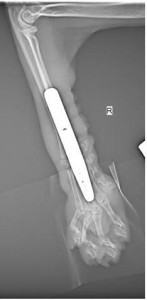
The carpus and manus possess an additional and permanent external rotation in relation to the antebrachium, which causes the patients to use the rostral portion of the their carpus for stepping and weight bearing, which in turn inducts the formation of a chronical traumatic inflammatory proliferative granuloma in this area. For about a 25-45 days period, an impossible to overcome carpal hyperfelexion develops, to the point where the joint can no longer be returned to its physiological position, due to the shortening of the flexor muscle-tendon apparatus (see video 1 with cat Sonia 39 days after the trauma:
A few therapeutic approaches are being advised for this pathologic condition worldwide: total limb amputation; stem cell therapy (with still controversial results); standard pancarpal arthrodesis (note that very often it is very difficult to execute procedure in the state of this disease and is almost always accompanied by a nonsatisfactory limb function end result).
None of the upper mentioned approaches for treatment of radial nerve palsy in cats, while trying to avoid limb amputation, was producing satisfying results in the patients with this problem, operated by our team. This is the reason we decided to test and implement a new “Dobro hrumvane modified pancarpal arthrodesis” procedure for the operative treatment of feline radial nerve palsy.
- Report patients base
Up to this moment, this modified by our team procedure has been done in 111 patients. In the first 11 patients we tried different but very similar to each other versions of the modification, and after patient 12 up to patient 111 (meaning exactly 100 patients) we were performing always one same version of the technique.
In 87 of these patients a follow-up postoperative monitoring for over one year has been performed (in 9 of them an approximately 5 year follow up was achieved, in 33 patients the follow up period was approximately 4 years etc.), in 11 patients the follow up period was between 4 months and one year and in 2 patients the follow up period was less than 4 months. In four of the operated patients, pre- or postoperative clinically relevant paralysis of the elbow region was also observed. As was mentioned earlier, 25% of feline radial nerve palsy patients demonstrate this (according to our observations in 23% of the patients it is already observed in the preoperative period and in other 2%, it develops a few weeks after the surgical intervention, with the reasons for that still being unclear). It should be noted that the majority of owners of patients with elbow area involvement preferred amputation over the experimental procedure.
- Surgical technique
The standard pancarpal arthrodesis general guidelines are being followed, but with the following modifications:
- Straight 11̊ inclination non locking hybrid pancarpal arthrodesis plate has been used (produced by Medimetal or Mikromed, delivered by VetWest). The plate contouring should be modified before the surgery and the inclination should become 21-22̊. Twisting of the distal portion of the plate internally in relation to the proximal portion of the plate is not recommended! For the fixation to the metacarpus 1.5 mm non locking screws were used (produced by Mikromed, delivered by VetWest) and for the fixation to the radius 2.0 mm non locking screws were used (produced by Mikromed, delivered by VetWest);
- The proximal (os carpi radiale et ulnare) and distal carpal bones are being completely removed, this being done with extreme caution not the traumatize the adjacent magistral structures (especially blood vessels), which are located on the palmar surface;
- The proximal ends of the metacarpal bones are being separated from one another;
- The fixation of the plate to the dorsal surface of the third metacarpal bone is achieved the same way as in the standard technique, using 1.5 mm thick and 6 mm long screws, but the fixation to the radius is not applied on its dorsal, but on its medial/mediocaudal edge/surface, using 2.0 mm screws. The screw hole on the plate which is intended for os carpi radiale (note that this bone is actually removed in the modified technique) is used for an additional 2.0 mm screw, placed in the distal radius. In other words, the whole metacarpal portion of the limb is being internally rotated around 85-95° (for the purpose of that an almost full blunt and careful separation of all soft tissues, including the magistral vessels and nerves in the distance between the carpus and the middle portion of the metacarpal bones, should be performed). After plating of the third metacarpal plate with four 1.5 mm non locking screws in neutral position the third metacarpal bone is being compressed to the radial distal This compression is easily achieved with the first screw, placed in the radius (not dorsal but medial/mediocaudal radial edge/surface – see below Xray picture Standard) thanks to the DC wholes of the plate types mentioned upper above. This screw is being inserted in the second 2.0 mm screw whole in distal to proximal direction, meaning the third plate hole in relation to the whole plate in proximal to distal direction. After that, 4 neutrally (not in compression mode) placed screws are applied to radial bone in the following order: the most distal hole, the most proximal hole, the second hole in proximal to distal direction, the third hole in proximal to distal direction. It is recommended that at least two of the screws in the distal radius engage the distal ulna too, so the distal portions of the two bones could eventually be pulled together – the screws could be numbers one and two or four and five from proximal to distal, this possibility could be estimated only intraoperativelly;
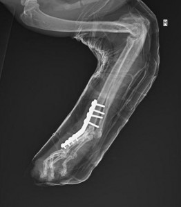
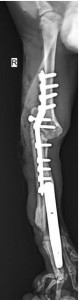
POST-OP STANDARD X-RAY PICTURE AFTER PANCARPAL ARTHRODESIS “DOBRO HRUMVANE”
- With this technique it is easy and recommendable to use a significant amount of autograft material – recommendable due to the large gap that is being created. This autograft is readily available, considering the amount of bone that is being removed in the previous stages of the surgical technique;
- The final stage of the surgery includes almost full blunt separation of the skin from the underlying soft tissues in the designated area, along with skin plastic traction modification, which is intended to place the fifth finger in a more medio-cranial position. The skin sutures and respectively the skin incision should be placed in a position that is not exactly above the plate (eventually they plate and incision could be placed in a cross manner, but should not be on top of each other for their whole lenght). It is not necessary to perform tenodesis of the digital extensors or excision of some skin on the dorsal carpal area in orther to pull the fingers in extension. It shoud be noted that the upper mentioned skin traction used to “pull” the fifth finger in a more dorsal and medial direction (meaning that the fifth finger is placed adjasent to the dorsolateral, not solely lateral, surface of the fourth finger, under subtle tension that will not allow overlapping of the fifth finger) is extremely important because in some of the first patients, which underwent the still not perfected procedure, weeks to months after the surgery pressure necrosis developped in the fifth finger, which required further revison plastic surgeries.
- In patients that have a very wild temper and where it is not possible to achieve two week long cage rest, postoperative splint could be placed. If this is done, additional amount of cotton could be used to help achieve the upper mentioned mediocranial position of the fifth metacarpus and finger;
- NEO K-9 clinical formula is prescribed for a month and a two week long cage rest is done in more calm patients.
- C) Results – the last 100 cats (No 12 … No 111 made with identical technique) :
C1) 96 patients that did not have (according to our clinical opinion) involvement of the elbow region pre- or postoperatively:
– 95 patients with good limb geometry in stance and during walking, active involvement of the limb during walks and playing, owners completely content with the results 4 months up to 5 years after the surgery. 89 of these 95 patients had no postoperative complicatioons; 2 patients developed moderate postoperative infection that was easily treated; 2 patients demonstrated delayed healing of the surgical incision in the area above the plate (it took more than 5 weeks in both patients); 2 patients had delayed bone union, that took around 5 months to be completed;
– 1 patient demonstrated unsatisfactory to this point level of weigt bearing and limb usage during walk and play. It is understandable that the owner of this patient is not completely content with the results, but is unfortunately refusing implant removal and further diagnostic procedures;
– No cases with implant loosening, intra- or postoperative fracture, postoperative necrosis etc.;
C2) 4 patients with clinically relevant pre- or postoperative involvement and paralysis of the motor unit of the elbow joint:
– 1 patient without preoperative elbow problem, developed such around a month after the surgical intervetion and the problem was accompanied by the development of an additional low grade external rotation of the antebrachium in relation to the limb portion above the elbow. The main problem was presented by progressive loss of support of the ebow joint in extension during stance, which lead to the inability of the limb to support the body during weight bearing. The problem was resolved after a two week long active rehabilitation and machine physiotherapy and application of a light splint, which is suporrting (but not blocking) the elbow.
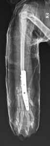
– 1 patient (cat named Trun) with preoperative paralysis of the elbow joint, but accompanied by almost complete ankylosis of the elbow joint (only 15% of the normal range of motion was preserved, especially the extension was blocked) – see below the post-op pictures of cat Trun
-
-
Although there was a serious accompanying problem, months after the surgery the owners are completely satisfied with the result. The patient is using the limb with no limitations during play, almost no limitations while running and with some limitations while walking – that last limitation is probably due to the constant flexed position of the elbow, which is exceeding the normal flexion angle of an elbow joint during walk, thus the animal is placing the shoulder of the affected limb under the level of the shoulder of the unaffected limb, during weight bearing (see video with cat Trun approx 3 months post-op:
-
The owners do not report signs of pain. Even though it is not right to make conclusions only on the basis of a single patient, this case gives us hope that patients with radial nerve palsy in combination with complete or partial elbow joint ankylosis have the chance to avoid amputation of the limb.
– 1 patient with partial preoperative paralysis of the elbow joint which became more severe (around 50%) month after the surgery: the bones in the arthrodesis region achieved complete healing, but the elbow joint loses support during weight bearing, thus the animal is placing the shoulder of the affected limb under the level of the shoulder of the unaffected limb, during weight bearing. Due to this the ptient is weight bearing the limb not on its pads, but rather on the carpal palmar angle surface. Because of that a chronic nonhealing skin lesion developed in this area over the time, which is intermitently bleeding. Up to this point, the owners are content with the result and do not wish to start rehabilitation or agree to a revision surgery, but for our team this result is unsatisfactory and it requires additional surgical and/or physiotrepautical intervention;
– 1 patient (Doxy) wtihout preoperative involvement and paralysis of the elbow, which developed a progressive clinically relevant paralysis of the elbow a few weeks after surgery. This led not only to loss of support of the elbow joint during weight bearing, but also to constant progressing additional rotation of the antebrachium in relation to the humeral area.
-
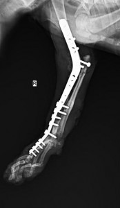
This rotation made the patient bear weight on the lateral surface of the carpal angle, developing a skin lesion there. This postoperative elbow joint paralysis did not resolve after a rehabilitation course. In order to correct the problem an elbow arthrodesis was performed, but not in a standard way. A “double-modified” elbow arthrodesis was performed: the boomerang plate produced by Mikromed and supplied by VetWest was placed on the lateral surface instead of the medial. Also, the antebrachial region was rotated 18 degrees internally, in relation to the humerus. We recommend very torough preoperative preparation: the execution of the technique is quite challenging, because the compression must be maintained and in the same time the “locking” of the anconeal process in the humeral fossa must be overcome, along with the congruency of the other ulnar structures and their corresponding radial structures – see below post-op X-ray pictures of cat Doxy after the second surgery, the elbow modified arthrodesis:
It can be seen that the plating is on the lateral surface of the radius distally and on the laterocranial surface of the humerus proximally.
Only a few hours after the surgery, the patient demonstrated excellent, pain free limb usage, with very good limb geometry and lack of difference in the level of the two shoulder joints during weight bearing. In the following days the patient started using the limb for playing too. At this point, 3-4 months after surgery, the patient is demonstrating completely satifying results (see video with cat Doxy approx 4 months post-op:
-
There are no signs of malunion, infection or other types of complications. The muscle mass in the shoulder area of the operated limb is similar to that of the non operated limb. Even if it is based just on one patient, the result of this case gives us some hope for surgical resolution for patients with modified or standard carpal arthrodesis, which have an acompanying or later develop severe elbow pathology of nonakylotic kind, as we know that the combination of carpal and elbow arthrodesis is not recommended in the known sources. For this patient especially we have an additional recommendation:
1) The first recommendation that is applied to all 111 operated patients – considering that it is a patient with a paralysed limb it should live on a non- smooth surface (but also not on an abrasive one). On a slippery surface patients with Dobro hrumvane arthrodesis step with mild slipping which combined with the lack of sensitivity could cause in longer period skin lesion (see Video 2 with cat Zhivka approx 5 weeks post-op:
-
2) Additional recommendation especially for Doxy: the patient has two joints that underwent arthodesis, which means that a stress point is being created between the two plates, which in turn creates a significant risk for further fractures. This risk is further amplified by the fact that the arthrodesis procedures are reducing the shock absorbing function of the joints. Considering all of the mentioned above, the patient should live in an enviornment that lacks the risk of creation of serious vertical vector forces (such as jumping to or from high places). It should be noted that Doxy did exactly that, many times after surgery and no problem occured, but it is still highly not recommended.
- Conclusion
The 100 clinical cases, with patients that underwent a similar modified pancarpal Dobro hrumvane arthrodesis procedure for the treatment of feline radial nerve palsy demonstrate a constant and satisfying result with very good return to function of the limb, pain free, with no discomfort. No following complications, including long-term ones are being observed and there is a very high level of owner satisfaction. We recommend this surgical technique and we would be glad to recieve feedback afer the completion of the procedure, either in the algorithm recommended by us, or with any additional modifications.
-
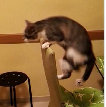
Even when the rotation of the metacarpal area in comparison to the antebrachial area is not 85-95 degrees the patients use the leg and the owners are satisfied but the leg geometry is in our opinion not good looking. Cat number 11, the last before the standartized 100 patients chain, named Hari is such a case, the rotation was 78-80 %, the operation was made approximately 5 years ago. As you can see at the videos made 4 years post-op the patient uses the left operated leg even during acrobatic jumping (see below picture Hari)
-
-
and active playing 4 years after surgery:
-
- E) Post scriptum
A few years ago we presented the technique and its results, based on a few dozens of cases, on a VOG\BAVOT event. Ever since, a few colleagues from the Balkan region have sent us feedback with very encouraging results, after using the technique. One of them was our inconsolable friend, colleague and inspirator, D-r V. Vasilev, whose memmory and collosal contribution to the development of the veterinary meidicine in Bulgaria we would like to honor in the end of this report.
-
Sofia The Orthopedic department of
March 2019 “Dobro hrumvane!” veterinary clinics